Anatomy Of Chest And Heart / Aorta Anatomy Function And Significance : Do you find the anatomy of the heart confusing?. Traditionally, the heart is described as having left heart and right heart chambers. Webmd's heart anatomy page provides a detailed image of the heart and provides information the heart has four chambers: Normal anatomy of the thorax on labeled chest ct: This chapter is an abbreviated review of thoracic anatomy as seen on chest radiographs and computed tomography. Our picks for anatomy of the heart and blood vessels.
Webmd's heart anatomy page provides a detailed image of the heart and provides information the heart has four chambers: This chapter is an abbreviated review of thoracic anatomy as seen on chest radiographs and computed tomography. Stable angina is the most common. It is located in the middle cavity of the chest, between the lungs. Compression of the heart and great vessels may cause murmurs.
X Ray Image Of The Chest Showing The Internal Anatomy Of The Stock Photo Picture And Royalty Free Image Image 81600412 from previews.123rf.com If we want to understand how the heart performs its vital role, we will first have to look at its structure, i.e., cardiac anatomy. Do you find the anatomy of the heart confusing? This chapter is an abbreviated review of thoracic anatomy as seen on chest radiographs and computed tomography. The right atrium and left atrium receive blood returning from the systemic and pulmonary circuits. The heart is a muscular organ in most animals, which pumps blood through the blood vessels of the circulatory system. Heart anatomy focuses on the structure and function of the heart. Radiological anatomy of the lungs, mediastinal lymph nodes, trachea, bronchi, pleural cavity, heart and pulmonary vessels. Normal thoracic ct (lungs, pleura, mediastinum and heart).
Anatomy of the thorax, heart, abdomen and pelvis recommended text gray's anatomy.
The loose fitting superficial part of this sac is the fibrous pericardium. Related online courses on physioplus. Do you find the anatomy of the heart confusing? The right atrium and left atrium receive blood returning from the systemic and pulmonary circuits. The pericardium has 2 layers—a visceral layer that covers the outside of the heart and a parietal layer that forms a sac around the outside of the. If we want to understand how the heart performs its vital role, we will first have to look at its structure, i.e., cardiac anatomy. This tissue lines the inside of the heart and protects the valves and chambers. Learn about the organ's amazing power and the functions of its many parts. Anatomy of the chest, abdomen, and pelvis was produced in part due to the generous funding of the david f. Narrowed coronary arteries cause predictable chest pain or discomfort with exertion. Located between the lungs in the middle of the chest, the heart pumps blood through the network of arteries and veins known as the cardiovascular system. Heart dissection gcse a level biology neet practical skills. Learn more about the heart in this article.
Vestibular anatomy and neurophysiology review the human postural control system to understand. Do you find the anatomy of the heart confusing? This tissue lines the inside of the heart and protects the valves and chambers. Your heart works as a pump that pushes blood to the organs, tissues, and cells of your body. Yen ho, phd frcpath fesc fhea royal brompton hospital.
Cg Image Of Woman S Chest Area Heart Major Arteries And Veins Stock Images Page Everypixel from media.istockphoto.com The heart is a muscular organ in most animals, which pumps blood through the blood vessels of the circulatory system. Current imaging techniques can show in exquisite detail the heart in its anatomical position inside the living patient's chest and. Do you find the anatomy of the heart confusing? Our picks for anatomy of the heart and blood vessels. Heart is a muscular organ sited in the mediastinum. The heart is located in the center of the chest with its apex toward the left. Radiological anatomy of the lungs, mediastinal lymph nodes, trachea, bronchi, pleural cavity, heart and pulmonary vessels. Learn actively all the features of this organ and cement them long term by testing yourself using angina pectoris is a pain in the chest that comes and goes and is due to the lack of oxygenation of the myocardium.
The conducting system of the heart.
Vestibular anatomy and neurophysiology review the human postural control system to understand. Vestibular anatomy and neurophysiology online course: ■ identify the basic anatomy seen on a chest radiograph. It has four hollow heart chambers surrounded by muscle and other heart tissue. Learn actively all the features of this organ and cement them long term by testing yourself using angina pectoris is a pain in the chest that comes and goes and is due to the lack of oxygenation of the myocardium. Heart functionally can be separated in left and right side. Your heart works as a pump that pushes blood to the organs, tissues, and cells of your body. The heart and circulatory system make up your cardiovascular system. The heart has two receiving chambers, and two pumping chambers. ■ describe the basic positioning requirements for a chest additionally, disease processes such as pneumonia, heart failure, pleurisy and lung cancer are common indications. Your heart is in the center of your chest, near your lungs. Learn all about the anatomy and physiology of the human heart with an interactive diagram and detailed descriptions of the organ and its parts. Traditionally, the heart is described as having left heart and right heart chambers.
Therefore, the funnel chest is also called 'cobbler chest'. Compression of the heart and great vessels may cause murmurs. Narrowed coronary arteries cause predictable chest pain or discomfort with exertion. The heart sits on the main muscle of breathing (the diaphragm), which is found beneath the lungs. This image shows the four chambers of the heart and the direction that blood flows through the heart.
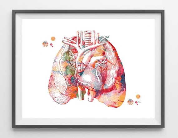
Lungs And Heart Anatomy Art Print The Heart In The Middle Of Etsy from i.etsystatic.com O heart—right ventricle, right ventricular outflow tract, left atrium, left ventricle, locations of the four cardiac valves. The heart is a muscular organ that pumps blood throughout the body. Stable angina is the most common. The heart is one of the most vital and delicate organs in the body. The heart and circulatory system make up your cardiovascular system. Vestibular anatomy and neurophysiology online course: Our picks for anatomy of the heart and blood vessels. Your heart does a lot of work to keep the body going.
Learn about the organ's amazing power and the functions of its many parts.
The loose fitting superficial part of this sac is the fibrous pericardium. The right atrium and left atrium receive blood returning from the systemic and pulmonary circuits. Narrowed coronary arteries cause predictable chest pain or discomfort with exertion. When a patient flexes the neck forward, the prominent process is usually that of the 7th cervical. The conducting system of the heart. Learn about the organ's amazing power and the functions of its many parts. The heart is a muscular organ that pumps blood throughout the body. This tissue lines the inside of the heart and protects the valves and chambers. Anatomy of the chest wall. Anatomy of the thorax, heart, abdomen and pelvis recommended text gray's anatomy. Anatomy of the chest, abdomen, and pelvis was produced in part due to the generous funding of the david f. Heart, organ that serves as a pump to circulate the blood. Traditionally, the heart is described as having left heart and right heart chambers.
Traditionally, the heart is described as having left heart and right heart chambers anatomy of chest. Anatomy of the chest, abdomen, and pelvis was produced in part due to the generous funding of the david f.


0 Komentar